The foot supports the body weight and provides leverage for walking and running. It is unique in that it is constructed in the form of arches, which enable it to adapt its shape to uneven surfaces. It also serves as a resilient spring to absorb shocks, such as in jumping. In anatomical terms, the foot is the pes. There are three parts of foot including the hindfoot, midfoot and forefoot. The top of the foot is the dorsum of the foot (or superior aspect). The bottom of the foot is the sole or plantar side or ventral side (or inferior aspect). The great toe (big toe) is digit #1 or the hallux while the small finger is digit# 5 or little finger of foot.
The Foot is Mainly Divided into Three Parts
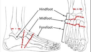
Forefoot, Midfoot and Hindfoot.
i. Forefoot
The forefoot is composed of 5 metatarsals, 14 phalanges and is present distal to the mid foot. The joints present in the forefoot are interphalangeal and metatarsophalangeal joints. These all structures belong to the distal parts of foot.
ii. Midfoot
The midfoot is composed of tarsal bones and three ligaments. It is pyramid in shape and connects the hindfoot to the forefoot. The mid parts of foot.
iii. Hindfoot
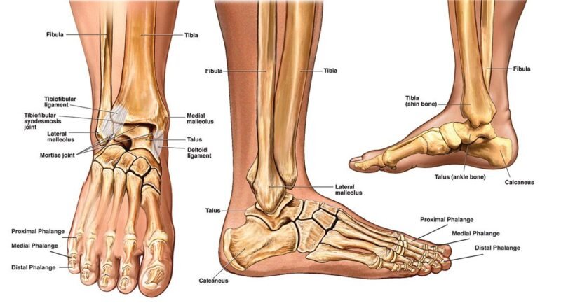
The hindfoot is composed of talus (ankle bone) and calcaneus (heel bone). These bones connect with tibia and fibula (Leg bones) to form the ankle joint. All these components are present in the proximal parts of foot.
Surfaces of Foot
While discussing the parts of foot lets discuss surfaces of the foot. The two surfaces of foot are as:
-
- Ventral surface (Sole or bottom of foot)
-
- Dorsal surface (Top surface of foot)
1. Ventral Surface/Sole of Foot
The ventral surface is the most important among the parts of foot. The major functions like walking, running, jumping are aided by the sole. The outermost layer (skin) of the sole of the foot is thick and hairless. It is the most exposed layer among parts of foot. Skin is firmly bound down to the underlying deep fascia by numerous fibrous bands. The skin shows a few flexural creases at the sites of skin movement. Sweat glands are present in large numbers.
a. Deep Fascia
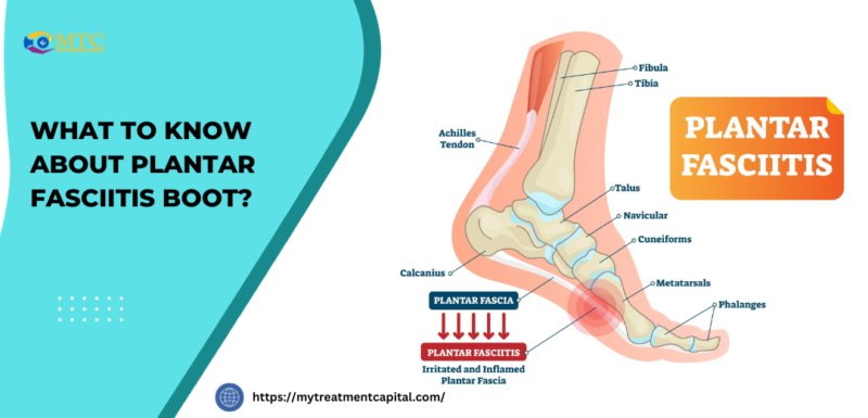
Plantar aponeurosis is a deep fascia thickening. It originates from the back of the foot (calcaneus bone) and extends till the toe. It is thick in the centre and thin at the sides of the foot. Plantar aponeurosis protects the deeper parts of foot including vessels, nerves and other underlying parts of foot. lt fixes the skin of the sole and helps in maintaining the longitudinal arches of the foot.
b. Sole Muscles
The muscles of the sole are conveniently described in four layers from superficial to deep.
i. First Layer
Abductor hallucis, flexor digitorum brevis, abductor digiti minimi.
ii. Second Layer
Quadratus plantae, lumbricals, flexor digitorum longus tendon, flexor hallucis longus tendon.
iii. Third Layer
Flexor hallucis brevis, adductor hallucis, flexor digiti minimi brevis.
iv. Fourth Layer
Interossei, fibularis longus tendon, tibialis posterior tendon.
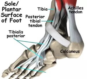
c. Long Tendons of Sole
Several muscles that originate in the posterior and lateral compartments of the leg send long tendons into the sole to their points of insertion. Following are some examples
-
- Flexor Digitorum Longus Tendon
-
- Flexor Hallucis Longus Tendon
-
- Fibrous Flexor Sheaths
-
- Synovial Flexor Sheaths
-
- Fibularis Longus Tendon
-
- Tibialis Posterior Tendon
All these long tendons help to maintain the movements in different parts of foot. The movements such as inversion, eversion, elevation, depression of foot are aided by these long tendons.
2. Dorsum of Foot
Dorsum of foot is composed of skin, muscles (Abductor digitimini, Flexor digitorum brevis) and top surface of bones of foot. The main artery for the dorsum of the foot is the dorsalis pedis artery. Dorsum of foot also helps in major functions like bearing weight, walking and running.
Arteries of Foot
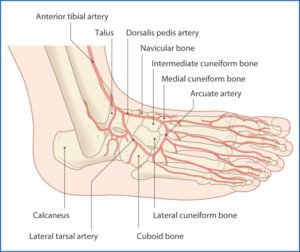
The muscles and certain other parts of foot are supplied by two arteries.
The dorsum of foot is supplied by dorsalis pedis artery while sole of foot is supplied by posterior tibial artery which passes behind the medial malleolus, and terminates by dividing into the medial and lateral plantar arteries.
i. Medial Plantar Artery
The medial plantar artery is the smaller of the terminal branches of the posterior tibial artery. It ends by supplying the medial side of parts of foot and the big toe. It gives off multiple muscular, cutaneous, and articular branches.
ii. Lateral Plantar Artery
The lateral plantar artery is the larger of the terminal branches of the posterior tibial artery. It also gives off numerous muscular, cutaneous, and articular branches along the lateral parts of foot.
Joints of Foot
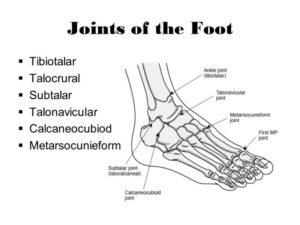
Functionally, there are three compound joints in the foot:
i. Subtalar or Talocalcaneal Joint
The clinical subtalar joint (synovial joint) between the talus and the calcaneus, where inversion and eversion occur about an oblique axis. The joint participates in the movements of inversion and eversion of the foot of all parts of foot.
ii. Transverse Tarsal Joint
The transverse tarsal joint, where the midfoot and forefoot rotate as a unit on the hindfoot around a longitudinal axis, helping in the inversion and eversion of foot. This is a ball and socket joint.
iii. Other Joints in Foot
Metatarsophalangeal and interphalangeal joints and certain plane joints involving the navicular, cuneiform, cuboid and the metatarsal bones. They involve elevation, depression, pronation and supination of the foot. All these joints are used to join different parts of foot.
Arches
A segmented structure can hold up weight only if it is built in the form of an arch. The foot has three such arches, which are present at birth: the medial longitudinal, lateral longitudinal, and transverse arches. In the young child, the foot appears to be flat because of the presence of a large amount of subcutaneous fat on the sole of the foot.
Types of Arches of Foot
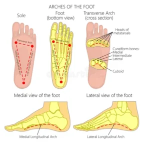
The arches provide the resilience necessary for walking, running, and jumping, and are maintained by four layers of passive, fibrous support, plus the dynamic support provided by the intrinsic muscles of the foot, and the long fibular, tibial, and flexor tendons.
i. Medial Longitudinal Arch
This consists of the calcaneus, the talus, the navicular bone, the three cuneiform bones, and the first three metatarsal bones among the parts of foot.
ii. Lateral Longitudinal Arch
This consists of the calcaneus, the cuboid, and the fourth and fifth metatarsal bones.
iii. Transverse Arch
This consists of the bases of the metatarsal bones and the cuboid and the three cuneiform bones.
iv. Integrity of the Bony Arches
The integrity of the bony arches of the foot is maintained by both passive factors and dynamic supports.
Passive factors involved in forming and maintaining the arches of the foot
- The shape of the united bones (both arches, but especially the transverse arch).
- Four successive layers of fibrous tissue that bowstring the longitudinal arch (superficial to deep)
- Plantar aponeurosis.
- Long plantar ligament.
- Plantar calcaneocuboid (short plantar) ligament.
- Plantar calcaneonavicular (spring) ligament.
Dynamic supports involved in maintaining the arches of the foot include.
-
- Active (reflexive) bracing action of intrinsic muscles of foot (longitudinal arch).
-
- Active and tonic contraction of muscles with long tendons extending into foot.
-
- Flexors hallucis and digitorum longus for the longitudinal arch.
-
- Fibularis longus and tibialis posterior for the transverse arch.
Conclusion
Foot is the major component of the human body that helps in mobility of mankind. The three parts of foot are hindfoot, midfoot and forefoot. Among all components of foot, arches of foot are of great importance that help to maintain the balance and bear weight while walking, running or jumping on uneven surfaces. Dorsum (upper) and Ventral (sole) of foot are the two surfaces of foot which are supplied by dorsalis pedis and posterior tibial artery respectively. Major movements of foot are inversion (Sole of foot facing towards the midline/inward), eversion (sole of foot facing outward), elevation and depression.